
The HELIOS Confocal Microscope is developed for transient absorption microscopy of a wide variety of objects. The microscope can be set up independently or as an add-on to an existing HELIOS ultrafast transient absorption spectrometer. The unique design enables measurements with sub-2 um spatial resolution. Users can switch between the reflection and transmission modes when studying thin films, semiconductor wafers, nanostructures, etc. This microscope enables studying micron-size samples or specific areas on heterogeneous samples. The built-in 2D camera makes it easy to find a spot of interest.
Spectrally resolved microscopy and spatially resolved spectroscopy measurements. The measurement is performed with a white light probe, a wide-range (450-750 nm) high-resolution (4 nm) spectrum is obtained instantaneously without the necessity to scan the probe wavelength.
Support for large samples. Long working distance objective lenses are used. A 22 mm-long sample area is available under transmission mode. Large space can handle thick substrates and makes loading the sample easy.
Direct viewing of the sample. The sample is illuminated by a dedicated LED. A color 2D camera collects the light and forms an image of the magnified sample through the objective lens. It makes finding an interesting sample area easy.
Tracking of the measured spot. The laser beams and the sample view can be sent to the camera to determine the location and spot size precisely.
Precise sample position control. Differential micrometers on an XYZ stage enable the control of a measured spot location.
The reflection mode facilitates the measurement of opaque or thick samples.
Simplified switching between transmission and reflection modes.
Customizable optical setup.
Spot Size
< 2 μm
Imaging Resolution
1.5 μm (for 750 nm)
Spectral Range
450 – 750 nm
Time Resolution
~300 fs (with a 70 fs laser pulse)
White Light Pulse Duration (Chirped Duration)
7 ps
Camera Viewing Window
68 µm x 90 μm
Objective Lens Numerical Aperture
0.6
Objective Working Distance
11 mm
Magnification
50X
Camera Imaging
Color
Reflection Mode
Optional

The image of MoS2 monolayer triangles on sapphire substrate is captured by the camera associated with the transient absorption microscope. Within the same image, the position of the focused pump and probe light is also shown. The center of circular target in the image marks a conjugated position with the entrance of the detector fiber.
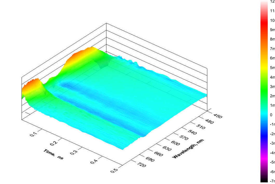
3D surface of measurement of MoS2 monolayer triangles with transient absorption microscope.
The measurement is conducted under the transmission mode of the microscope.

3D surface of measurement of MoS2 monolayer triangles with transient absorption microscope.
The measurement is conducted under the transmission mode of the microscope.
This site is protected by reCAPTCHA and the Google Privacy Policy and Terms of Service apply.
© 2002 – 2024 Ultrafast Systems, 8330 Consumer Ct, Sarasota, FL 34240, USA. All Rights Reserved.
An answer to your question might already be there.